
Doppler Ultrasound: Vascular Imaging at Sanconfind Medical Center
Blood flow is essential to life, delivering oxygen and nutrients to every tissue in the body. When blood flow is compromised by arterial blockages, venous clots, or other vascular conditions, serious health consequences can follow. Doppler ultrasound provides a non-invasive way to visualize and measure blood flow throughout the body, helping diagnose vascular conditions before they cause significant harm.
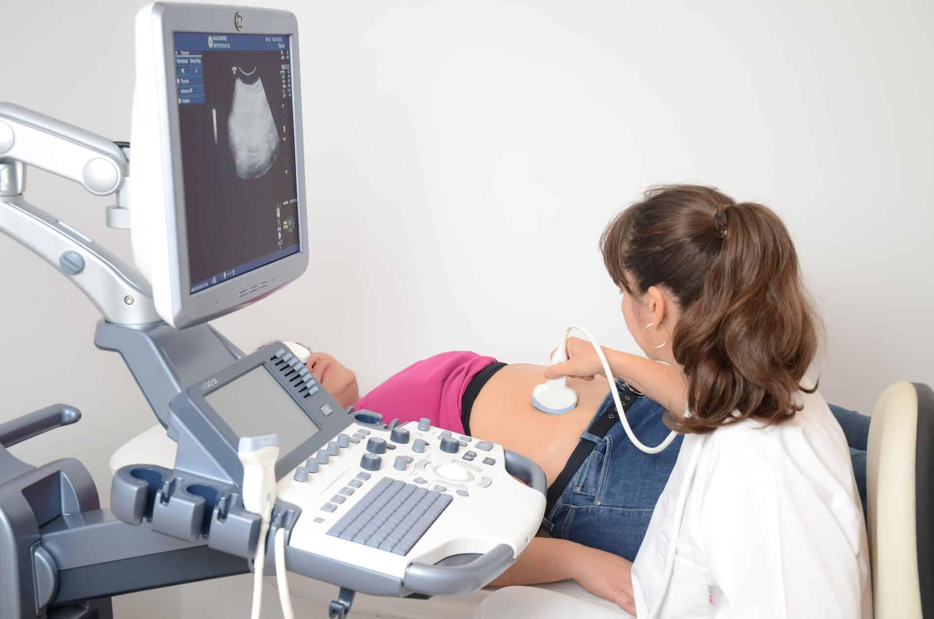
At Sanconfind Medical Center in Poiana Câmpina, Romania, our Doppler ultrasound services enable comprehensive vascular assessment without radiation exposure, supporting the diagnosis and management of conditions affecting arteries and veins throughout the body.
Understanding Doppler Ultrasound
The Doppler Principle
Doppler ultrasound is based on the Doppler effect—the same phenomenon that causes a siren to sound higher-pitched as an ambulance approaches and lower-pitched as it moves away. In medical imaging, this principle is applied to detect blood flow.
How It Works
- Ultrasound waves are transmitted into blood vessels
- Moving blood cells reflect the waves back
- The frequency shift of returning waves indicates blood movement
- Direction and speed of flow can be determined
- Results are displayed visually and as waveforms
Types of Doppler
Color Doppler
- Shows blood flow as color overlaid on grayscale images
- Red typically indicates flow toward the transducer
- Blue indicates flow away from the transducer
- Quick visual assessment of flow presence and direction
Power Doppler
- More sensitive than color Doppler
- Detects low-velocity flow
- Doesn't show direction—just presence of flow
- Useful for small vessels and low-flow states
Spectral (Pulsed-Wave) Doppler
- Displays flow characteristics as waveforms
- Shows velocity over time
- Allows precise measurements
- Reveals flow patterns characteristic of different conditions
Duplex Ultrasound Combines standard B-mode imaging with Doppler—the approach used most commonly in vascular examinations.
Doppler Ultrasound Applications
Doppler technology enables comprehensive vascular assessment across multiple body regions.
Carotid Doppler
Why It's Important The carotid arteries supply blood to the brain. Narrowing (stenosis) due to atherosclerosis can lead to stroke—a leading cause of death and disability.
What It Evaluates
- Common, internal, and external carotid arteries
- Vertebral arteries
- Degree of narrowing (stenosis)
- Plaque characteristics
- Blood flow velocities
Indications
- Transient ischemic attack (TIA) or stroke
- Carotid bruit (abnormal sound heard with stethoscope)
- Pre-surgical cardiovascular evaluation
- Follow-up after carotid procedures
- Screening in high-risk patients
Venous Doppler: Lower Extremity
Deep Vein Thrombosis (DVT) Detection DVT—blood clots in the deep leg veins—is a serious condition that can lead to pulmonary embolism (PE), a potentially fatal complication.
What It Evaluates
- Common femoral vein
- Femoral and deep femoral veins
- Popliteal vein
- Calf veins (when indicated)
- Vein compressibility (clots prevent compression)
- Flow augmentation and spontaneity
Indications
- Leg swelling, pain, or redness
- Risk factors for DVT
- Pre-operative screening
- Follow-up of known DVT
- Evaluation before treatment
Venous Doppler: Upper Extremity
Applications
- Suspected arm DVT
- Catheter-related thrombosis
- Pre-dialysis access planning
- Swelling evaluation
Arterial Doppler: Lower Extremity
Peripheral Artery Disease (PAD) Atherosclerosis affecting leg arteries causes claudication (leg pain with walking) and can progress to critical limb ischemia.
What It Evaluates
- Femoral arteries
- Popliteal arteries
- Tibial and pedal arteries
- Degree of stenosis
- Flow patterns
- Ankle-brachial index (ABI)
Indications
- Claudication (leg pain with walking)
- Non-healing leg or foot wounds
- Absent pulses
- PAD risk factor assessment
- Pre-surgical evaluation
Renal Doppler
Renal Artery Stenosis Narrowing of arteries supplying the kidneys can cause hypertension and kidney damage.
What It Evaluates
- Renal artery flow velocities
- Resistive indices
- Kidney size and appearance
- Post-procedure assessment
Indications
- Resistant hypertension
- Renal artery stenosis screening
- Kidney transplant evaluation
- Unexplained kidney dysfunction
Abdominal Vascular Doppler
Applications
- Portal vein evaluation (liver disease)
- Hepatic vein assessment (Budd-Chiari syndrome)
- Mesenteric artery evaluation
- Aortic aneurysm flow assessment
Transcranial Doppler
Intracranial Blood Flow Evaluates blood flow in major brain arteries through the skull.
Applications
- Stroke risk assessment
- Vasospasm monitoring
- Sickle cell disease screening
- Brain death evaluation
Obstetric Doppler
Fetal and Maternal Assessment
- Umbilical artery Doppler (fetal well-being)
- Middle cerebral artery flow
- Uterine artery Doppler
- Ductus venosus assessment
The Doppler Examination Experience
Before Your Examination
Preparation Most Doppler examinations require minimal preparation:
- Carotid Doppler: No special preparation
- Abdominal vascular: May require fasting
- Lower extremity venous: Usually no preparation
- Wear comfortable, loose clothing
We'll provide specific instructions when scheduling.
During the Examination
The Procedure
- You'll be positioned appropriately for the examination
- Warm gel is applied to the skin
- The sonographer places the transducer on your skin
- Images and Doppler waveforms are recorded
- Measurements are taken at specific locations
- The exam takes 30-60 minutes depending on the study
What You'll Experience
- Pressure from the transducer
- Possible compression of veins (checking for compressibility)
- Audible sounds from blood flow (Doppler signal)
- The examination is painless
After the Examination
Results
- Images and measurements are reviewed
- A physician interprets the findings
- Report is sent to your referring doctor
- Urgent findings are communicated promptly
Vascular Conditions Detected
Arterial Conditions
Atherosclerosis Plaque buildup in artery walls, leading to:
- Carotid stenosis (stroke risk)
- Peripheral artery disease (leg symptoms)
- Renal artery stenosis (hypertension)
Aneurysms Abnormal artery enlargement, especially:
- Abdominal aortic aneurysm
- Popliteal aneurysm
Dissection Arterial wall separation (emergency condition)
Venous Conditions
Deep Vein Thrombosis (DVT) Blood clots in deep veins—a medical emergency due to PE risk
Chronic Venous Insufficiency Venous valve failure causing:
- Leg swelling
- Varicose veins
- Skin changes
Superficial Thrombophlebitis Clots in superficial veins (usually less serious but may need treatment)
Why Choose Sanconfind for Doppler Ultrasound?
Advanced Technology
Our Doppler capabilities include:
- High-resolution imaging
- Color and power Doppler
- Spectral analysis
- Measurement and calculation tools
- Image archiving
Experienced Team
Our vascular imaging is performed by:
- Trained vascular sonographers
- Physician oversight
- Quality-focused protocols
- Ongoing education
Comprehensive Vascular Care
Doppler findings integrate with:
- Our phlebology services (vein treatment)
- Cardiology consultations
- Surgical referral when needed
- Follow-up imaging
Patient-Centered Service
We provide:
- Convenient scheduling
- Comfortable examination environment
- Clear communication
- Prompt reporting
Frequently Asked Questions
Is Doppler ultrasound the same as regular ultrasound?
Doppler ultrasound adds blood flow assessment to standard imaging. Regular ultrasound shows anatomy; Doppler shows moving blood. Most vascular exams combine both (duplex ultrasound).
Is Doppler ultrasound safe?
Yes. Like all ultrasound, Doppler uses sound waves—no radiation. It's safe for all patients, including pregnant women.
Does Doppler ultrasound hurt?
No. You'll feel pressure from the transducer and possibly some compression when checking for vein compressibility, but the exam isn't painful.
How accurate is Doppler for detecting blood clots?
Doppler ultrasound is highly accurate for detecting DVT in the thigh and above the knee. Accuracy is good but somewhat lower for calf vein clots.
Can Doppler tell how blocked an artery is?
Yes. Flow velocities and waveform patterns indicate the degree of arterial narrowing. We can estimate percentage stenosis.
Do I need to prepare for a Doppler exam?
Usually minimal or no preparation is needed. We'll provide specific instructions based on which examination you're having.
When will I get results?
Results are typically available within 24-48 hours. Urgent findings are communicated immediately to your doctor.
Is Doppler better than CT or MRI for vascular imaging?
Each has advantages. Doppler is non-invasive, radiation-free, and shows real-time flow. CT and MRI provide different anatomical details. Your doctor recommends the appropriate test.
Schedule Your Doppler Examination
For accurate vascular assessment without radiation, contact Sanconfind Medical Center for Doppler ultrasound.
Contact Us
- Phone: +40 244 990
- Email: office@sanconfind.ro
- Location: Sanconfind Medical Center, Poiana Câmpina, Prahova County, Romania
Our team will answer questions, provide preparation instructions, and schedule your examination.
Sanconfind Medical Center's Doppler ultrasound services provide comprehensive vascular assessment, supporting the diagnosis and management of conditions affecting arteries and veins throughout the body.