Ultrasound: Safe Diagnostic Imaging at Sanconfind Medical Center
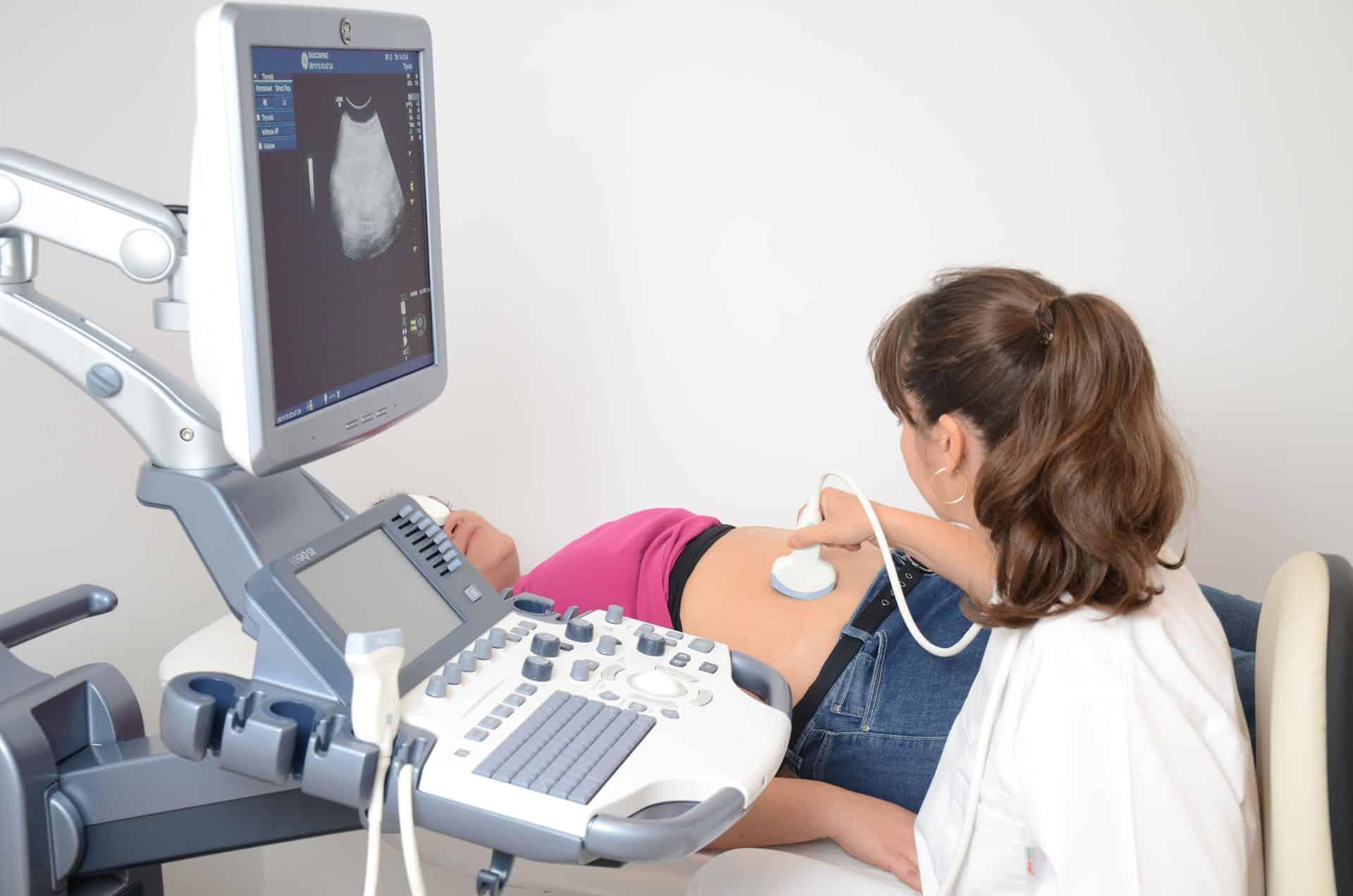
Ultrasound imaging, also known as sonography, uses high-frequency sound waves to create images of internal body structures. This versatile, safe, and non-invasive technology plays a crucial role in modern medical diagnosis, from monitoring pregnancy to detecting tumors and guiding medical procedures. At Sanconfind Medical Center in Poiana Câmpina, Romania, our ultrasound services provide patients with high-quality diagnostic imaging without radiation exposure.
Our skilled sonographers and physicians use advanced ultrasound equipment to visualize organs, tissues, and blood flow, supporting accurate diagnosis and effective treatment planning across a wide range of medical conditions.
Understanding Ultrasound
How Ultrasound Works
Sound Wave Technology Ultrasound uses sound waves at frequencies too high for human hearing (typically 2-18 MHz). A transducer (probe) sends these waves into the body and receives the echoes that bounce back from internal structures.
Image Creation
- Sound waves penetrate the body and reflect off tissues
- Different tissues reflect sound differently
- A computer processes the returning echoes
- Real-time images are created and displayed on a screen
- Images can be saved and measured
Why It's Safe
- No ionizing radiation
- No known harmful effects at diagnostic levels
- Safe for pregnancy
- Can be repeated as often as needed
- Suitable for children and sensitive populations
Types of Ultrasound
B-Mode (2D) Ultrasound The most common type, creating two-dimensional grayscale images. Ideal for examining organs and structures.
Doppler Ultrasound Evaluates blood flow by detecting movement. Shows direction and speed of blood in vessels. (See our dedicated Doppler ultrasound service for more details.)
3D/4D Ultrasound Creates three-dimensional images (3D) or real-time 3D video (4D). Often used in obstetric imaging.
Elastography Assesses tissue stiffness, helpful for liver disease staging and characterizing masses.
Ultrasound Applications
Ultrasound serves diagnostic purposes across numerous medical specialties.
Abdominal Ultrasound
Liver
- Fatty liver disease
- Cirrhosis
- Liver masses and cysts
- Bile duct dilation
- Hepatitis assessment
Gallbladder
- Gallstones (primary diagnostic tool)
- Cholecystitis (inflammation)
- Polyps and masses
- Biliary system evaluation
Pancreas
- Pancreatitis
- Pancreatic masses
- Cysts and pseudocysts
- Ductal abnormalities
Kidneys
- Kidney stones
- Hydronephrosis (swelling)
- Masses and cysts
- Kidney size and structure
- Post-surgical evaluation
Spleen
- Size assessment
- Masses and cysts
- Trauma evaluation
Aorta
- Aneurysm screening
- Size measurement
Pelvic Ultrasound
Gynecological
- Uterine fibroids
- Ovarian cysts and masses
- Endometrial evaluation
- Pelvic inflammatory disease
- Infertility assessment
- IUD placement verification
Obstetric
- Pregnancy confirmation
- Fetal development monitoring
- Dating and growth assessment
- Anatomical survey
- Placental evaluation
- Amniotic fluid assessment
- Multiple pregnancy evaluation
Urological
- Bladder evaluation
- Post-void residual measurement
- Prostate assessment (transrectal)
- Scrotal examination
Thyroid and Neck Ultrasound
Thyroid Gland
- Nodule detection and characterization
- Thyroiditis evaluation
- Size assessment
- Guidance for biopsy
- Cancer surveillance
Other Neck Structures
- Lymph nodes
- Salivary glands
- Parathyroid glands
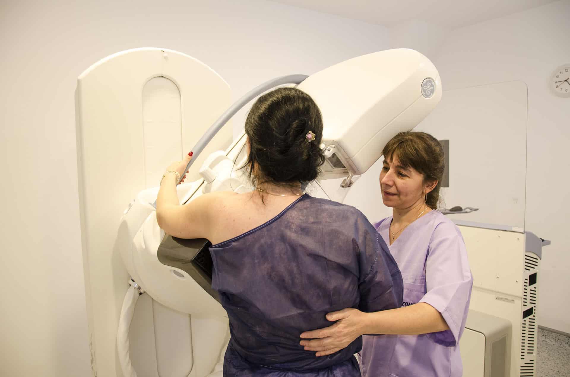
Breast Ultrasound
Applications
- Characterizing mammographic findings
- Evaluating palpable lumps
- Dense breast supplemental screening
- Guiding biopsy procedures
- Young patients with breast concerns
Musculoskeletal Ultrasound
Soft Tissue Evaluation
- Tendon injuries and inflammation
- Muscle tears and hematomas
- Ligament injuries
- Bursitis
- Joint effusions
- Nerve entrapment
- Foreign body detection
Advantages
- Dynamic imaging (can watch structures move)
- Comparison with opposite side
- Cost-effective alternative to MRI for some conditions
Vascular Ultrasound
Applications (see also Doppler ultrasound service)
- Deep vein thrombosis (DVT)
- Varicose vein assessment
- Arterial disease
- Carotid artery evaluation
Pediatric Ultrasound
Safe for Children
- Appendicitis (first-line imaging in children)
- Pyloric stenosis
- Intussusception
- Hip dysplasia screening
- Scrotal evaluation
- Renal anomalies
The Ultrasound Experience
Our ultrasound service is designed for patient comfort and diagnostic accuracy.
Before Your Examination
Preparation Varies by Study
Abdominal ultrasound:
- Typically requires fasting for 6-8 hours
- Allows gallbladder to fill and reduces bowel gas
Pelvic ultrasound (transabdominal):
- Full bladder required
- Drink water and don't urinate before the exam
Thyroid/neck/extremity ultrasound:
- Usually no special preparation
We'll provide specific instructions when you schedule.
During the Examination
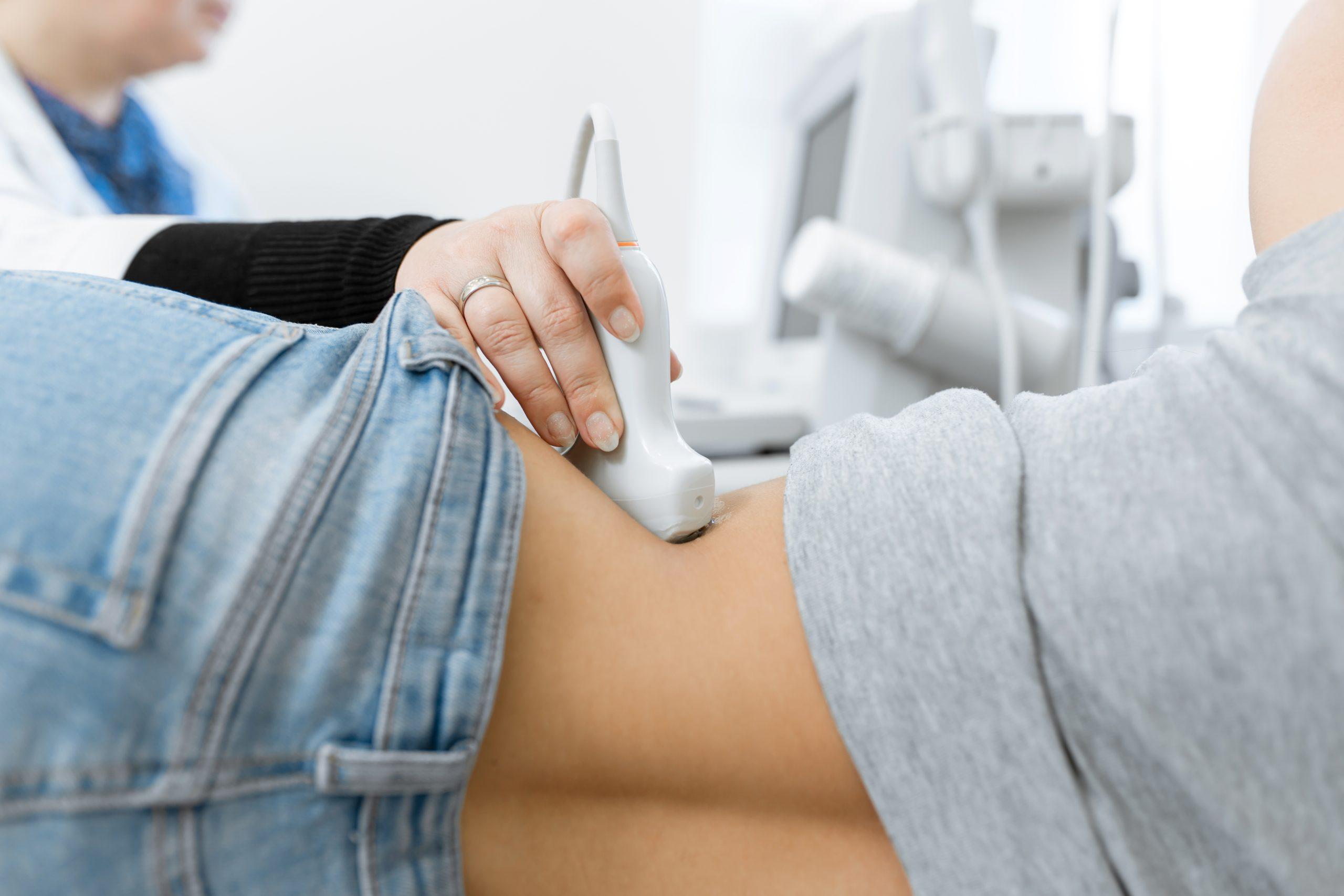
The Procedure
- You'll lie on an examination table
- Warm gel is applied to the skin (helps transmit sound waves)
- The sonographer presses the transducer against your skin
- The transducer is moved across the area of interest
- Images are captured and stored
- The exam takes 20-45 minutes depending on the study
What You'll Experience
- Pressure from the transducer (usually comfortable)
- Warm gel on your skin
- Possible request to hold your breath briefly
- You may see images on the screen
- Painless examination in most cases
After the Examination
Results
- Gel is wiped from your skin
- You can resume normal activities immediately
- Images are reviewed by a physician
- Formal report is provided to your referring doctor
- Emergency findings are communicated promptly
Why Choose Sanconfind for Ultrasound?
Modern Equipment
Our ultrasound systems feature:
- High-resolution imaging
- Multiple specialized transducers
- Doppler capabilities
- Image storage and archiving
- Measurement and analysis tools
Skilled Professionals
Our team provides:
- Experienced sonographers
- Physician interpretation
- Patient-focused care
- Technical expertise
- Quality examinations
Comprehensive Service
We offer:
- Wide range of examination types
- Convenient scheduling
- Integration with other services
- Prompt reporting
- Follow-up coordination
Patient Comfort
We emphasize:
- Warm, comfortable environment
- Privacy and dignity
- Clear communication
- Minimal wait times
- Professional, caring staff
Frequently Asked Questions
Is ultrasound safe?
Yes. Ultrasound uses sound waves, not radiation. It has an excellent safety record and is used safely throughout pregnancy and in patients of all ages.
Does ultrasound hurt?
No. Most ultrasound examinations are painless. You'll feel pressure from the transducer, which is usually comfortable. Some examinations may cause mild discomfort from a full bladder or probe pressure.
How long does an ultrasound take?
Most examinations take 20-45 minutes. Complex studies may take longer.
Do I need to prepare for ultrasound?
Preparation depends on the examination. Abdominal ultrasound usually requires fasting. Pelvic ultrasound often requires a full bladder. We'll provide specific instructions.
Can ultrasound detect cancer?
Ultrasound can identify masses and characterize them based on their appearance. While it can suggest whether a mass is concerning, biopsy is often needed for definitive diagnosis.
When will I get my results?
Our physicians review images and create reports typically within 24-48 hours. Urgent findings are communicated immediately.
Is ultrasound better than CT or MRI?
Each modality has strengths. Ultrasound excels at real-time imaging, is radiation-free, and is ideal for certain applications. CT and MRI are better for other conditions. Your doctor recommends the appropriate test.
Can I see my baby during prenatal ultrasound?
Yes. We can often show you images during the examination, though our primary focus is completing the diagnostic study.
Schedule Your Ultrasound
Experience safe, high-quality ultrasound imaging at Sanconfind Medical Center. Contact us to schedule your examination.
Contact Us
- Phone: +40 244 990
- Email: office@sanconfind.ro
- Location: Sanconfind Medical Center, Poiana Câmpina, Prahova County, Romania
Our team will answer your questions, provide preparation instructions, and schedule your ultrasound at a convenient time.
Sanconfind Medical Center's ultrasound services combine advanced technology with skilled professionals to provide safe, accurate diagnostic imaging without radiation exposure.