Mammography: Breast Cancer Screening at Sanconfind Medical Center
Breast cancer is one of the most common cancers affecting women worldwide, but early detection dramatically improves treatment success and survival rates. Mammography remains the gold standard for breast cancer screening, capable of detecting tumors before they can be felt during physical examination. At Sanconfind Medical Center in Poiana Câmpina, Romania, our mammography service provides women with high-quality breast imaging in a comfortable, supportive environment.
Our commitment to women's health includes not just excellent imaging technology, but also compassionate care that recognizes the anxiety many women feel about breast screening. We're here to support you through every step of the process.
Understanding Mammography
What is Mammography?
Mammography is a specialized X-ray examination of the breast designed to detect breast cancer and other breast abnormalities. Using low-dose X-rays, mammography creates detailed images of breast tissue that radiologists analyze for signs of cancer.
How Mammography Works
The Technology
- Specialized X-ray unit designed for breast imaging
- Compression paddles flatten the breast for optimal imaging
- Low-dose X-rays pass through breast tissue
- Digital detectors capture the image
- Computer processing enhances image quality
Why Compression is Necessary Breast compression during mammography:
- Spreads tissue to prevent overlapping structures
- Creates uniform breast thickness
- Reduces radiation dose needed
- Improves image quality
- Reduces motion blur
While compression is uncomfortable, it lasts only seconds per image.
Digital Mammography
Modern digital mammography (our technology) offers advantages over older film mammography:
- Immediate image availability
- Ability to adjust brightness and contrast
- Computer-aided detection capabilities
- Easier storage and retrieval
- Potentially lower radiation doses
- Better visualization of dense breast tissue
Types of Mammography
Screening Mammography
Purpose Routine examination of women without symptoms to detect breast cancer early.
Standard Views
- Craniocaudal (CC): Top-to-bottom view
- Mediolateral oblique (MLO): Angled side view
- Two views of each breast
Who Should Be Screened Guidelines vary, but generally recommend:
- Women 50-74: Screening every 1-2 years
- Women 40-49: Discuss with doctor based on risk factors
- High-risk women: May begin earlier, more frequently
Diagnostic Mammography
Purpose Evaluate symptoms or investigate abnormalities found on screening.
Additional Views
- Spot compression: Focuses on specific area
- Magnification: Enlarges suspicious region
- Special angles: Additional perspectives
- More images than screening
Indications
- Abnormal screening mammogram follow-up
- Breast lump or thickening
- Nipple discharge
- Breast pain
- Skin changes
The Mammography Experience
Understanding what to expect can ease anxiety about mammography.
Before Your Mammogram
Scheduling
- Best timing: 1-2 weeks after menstruation (breasts less tender)
- Bring prior mammogram images if from another facility
- Inform us of breast implants
Preparation
- Don't use deodorant, antiperspirant, powder, or lotion on breast/underarm area (can appear as artifacts on images)
- Wear two-piece clothing for convenience
- Report breast symptoms to technologist
Information to Provide
- Breast symptoms or concerns
- Previous breast procedures or surgeries
- Family history of breast cancer
- Hormone use
- Date of last menstrual period
During the Examination
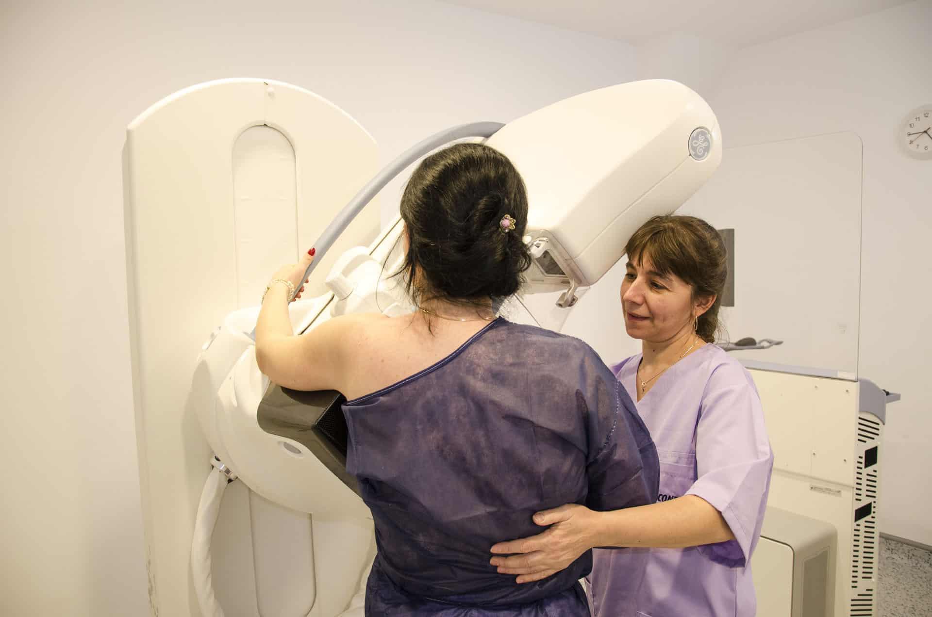
The Procedure
- You'll change into a gown from the waist up
- The technologist positions your breast on the imaging platform
- A compression paddle gently flattens the breast
- You'll hold your breath briefly while the image is taken
- Compression is released immediately after each image
- Process is repeated for different views and both breasts
Duration The examination takes approximately 15-30 minutes.
Discomfort Compression causes pressure and may be uncomfortable, but:
- Each compression lasts only seconds
- It's necessary for quality images
- Technologists work to minimize discomfort
- Communicate if you're in pain
After the Mammogram
Results
- Radiologists review your images
- Results are typically available within a few days
- You'll be contacted with findings
- If abnormalities are found, additional testing may be recommended
What Results Mean
- Negative: No abnormality detected
- Benign finding: Non-cancerous abnormality identified
- Probably benign: Very likely non-cancerous; short-term follow-up recommended
- Suspicious: Additional testing needed
- Highly suspicious: Biopsy recommended
Understanding Breast Density
What is Breast Density?
Breast density refers to the amount of fibroglandular tissue relative to fatty tissue in the breast. Dense breasts have more fibroglandular tissue, which appears white on mammograms—the same as tumors.
Why Density Matters
Imaging Challenge Dense tissue can hide cancers (masking effect), potentially reducing mammography sensitivity.
Cancer Risk Women with dense breasts have slightly higher breast cancer risk.
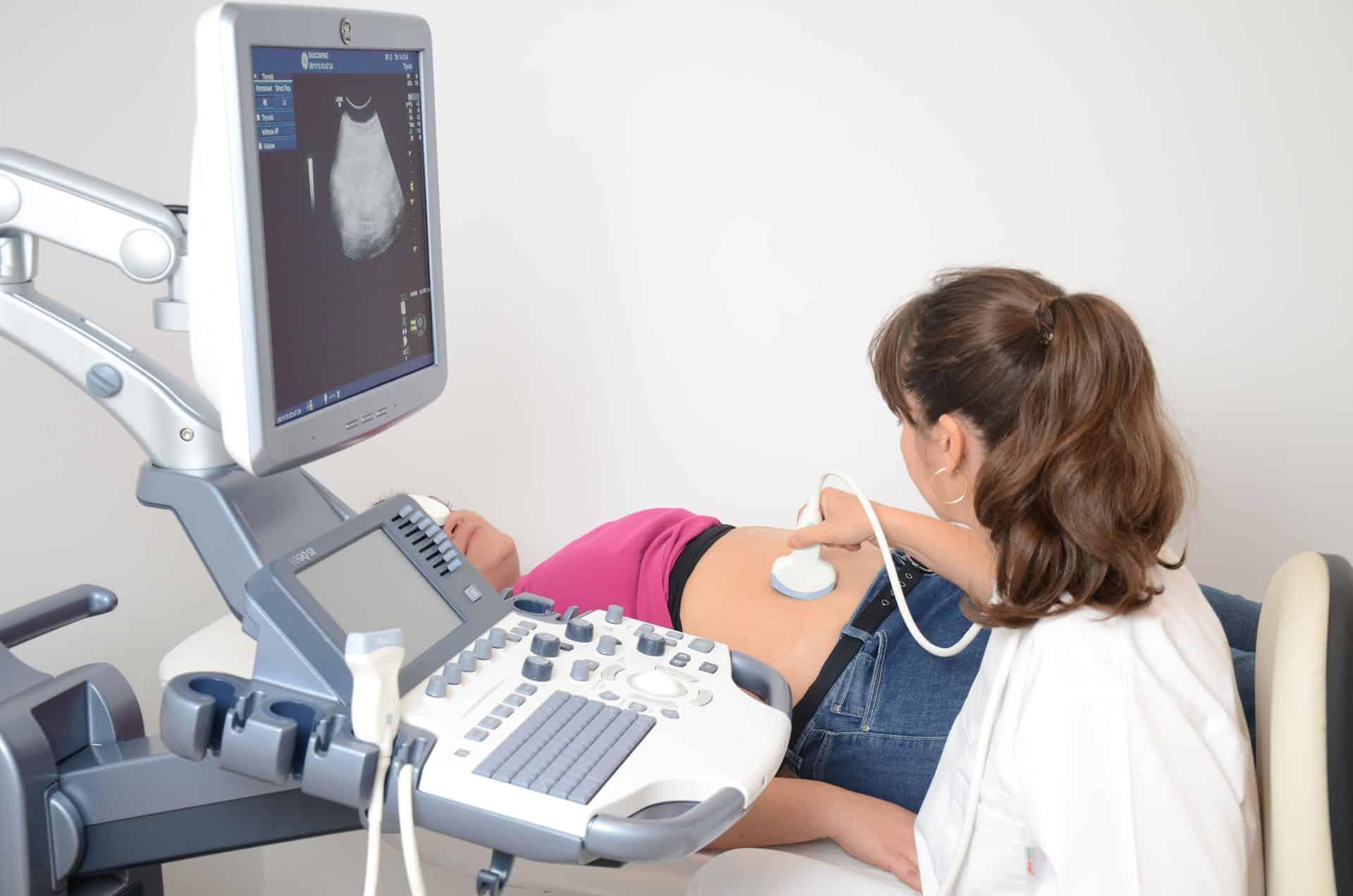
Supplemental Screening Women with dense breasts may benefit from additional imaging:
- Breast ultrasound
- Breast MRI (for high-risk women)
We'll inform you of your breast density and discuss options if appropriate.
Breast Cancer Risk Factors
Understanding risk helps determine screening approach.
Factors You Can't Change
- Age: Risk increases with age
- Family history: Especially first-degree relatives
- Genetic mutations: BRCA1, BRCA2
- Personal history: Previous breast cancer or certain benign conditions
- Dense breasts
- Reproductive factors: Early menstruation, late menopause, first pregnancy after 30
Factors You May Influence
- Obesity: Especially after menopause
- Physical inactivity
- Alcohol consumption
- Hormone therapy: Long-term combined HRT
- Not breastfeeding
Mammography and Early Detection
Why Screening Matters
Cancer Statistics
- Breast cancer is highly treatable when caught early
- 5-year survival for localized breast cancer exceeds 99%
- Mammography can detect cancers 1-3 years before they're palpable
- Regular screening reduces breast cancer mortality
What Mammography Detects
- Masses (tumors)
- Calcifications (tiny calcium deposits that may indicate cancer)
- Architectural distortion
- Asymmetries
- Other abnormalities
Limitations of Mammography
No test is perfect. Mammography may:
- Miss some cancers (false negatives), especially in dense breasts
- Find abnormalities that aren't cancer (false positives), leading to additional testing
- Detect cancers that might not have caused problems (overdiagnosis)
Despite limitations, mammography remains the best screening tool available and has saved countless lives.
Why Choose Sanconfind for Mammography?
Technology
Our mammography service features:
- Modern digital mammography equipment
- Low radiation dose
- High-resolution imaging
- Computer-aided detection tools
Expertise
Your mammogram is read by:
- Experienced radiologists
- Those trained in breast imaging
- Using systematic interpretation methods
- With access to prior studies for comparison
Patient Experience
We prioritize your comfort:
- Female technologists available
- Private, comfortable environment
- Compassionate care
- Clear communication
- Prompt results
Comprehensive Care
Mammography integrates with:
- Breast ultrasound when needed
- Gynecology services
- Surgical consultation if required
- Complete follow-up support
Frequently Asked Questions
How often should I have a mammogram?
Guidelines recommend screening every 1-2 years for women 50-74. Women 40-49 should discuss with their doctor. High-risk women may screen more frequently.
Is mammography safe?
Yes. Mammography uses very low radiation doses. The benefit of early cancer detection far outweighs the minimal radiation risk.
Does mammography hurt?
Compression is uncomfortable but brief—just seconds per image. Communicate with the technologist if you're in pain.
What if I have breast implants?
Mammography is possible with implants, but tell us beforehand. Special techniques ensure adequate breast tissue imaging while protecting implants.
Should I worry if I'm called back after screening?
Callbacks are common and usually lead to benign (non-cancer) diagnoses. Additional imaging provides clarity about areas that need closer look.
At what age should I stop mammography?
Discuss with your doctor. Many women continue screening into their 70s as long as they're in good health and would pursue treatment if cancer were found.
Does mammography detect all breast cancers?
No test detects all cancers. Mammography finds most breast cancers, but some (especially in dense breasts) may be missed. Report any breast changes to your doctor.
Schedule Your Mammogram
Take control of your breast health. Schedule your mammogram at Sanconfind Medical Center.
Contact Us
- Phone: +40 244 990
- Email: office@sanconfind.ro
- Location: Sanconfind Medical Center, Poiana Câmpina, Prahova County, Romania
Our team will schedule your appointment and answer any questions about breast imaging.
Sanconfind Medical Center's mammography service combines quality imaging technology with compassionate care, supporting the early detection of breast cancer and promoting women's health.