Digital Radiology: X-Ray Imaging at Sanconfind Medical Center
X-ray imaging, the oldest form of medical imaging, remains one of the most valuable diagnostic tools in medicine. From detecting bone fractures to evaluating lung conditions, X-rays provide rapid, accurate information that guides countless medical decisions daily. At Sanconfind Medical Center in Poiana Câmpina, Romania, our digital radiology services combine this proven technology with modern digital capabilities, delivering high-quality images with minimal radiation exposure.
Digital radiography offers significant advantages over traditional film X-rays, including immediate image availability, lower radiation doses, enhanced image quality, and easy sharing with other healthcare providers.
Understanding Digital Radiography
How X-Rays Work
The Technology X-rays are a form of electromagnetic radiation that can pass through the body. Different tissues absorb X-rays differently:
- Dense structures (bone, metal) absorb more X-rays and appear white
- Air-filled structures absorb very few X-rays and appear black
- Soft tissues appear in shades of gray
Digital vs. Film Traditional film X-rays captured images on photographic film. Digital radiography uses electronic detectors that convert X-rays directly into digital images, offering numerous advantages.
Advantages of Digital Radiography
Immediate Availability Images appear on the screen within seconds—no chemical development needed.
Image Enhancement Digital images can be:
- Adjusted for brightness and contrast
- Magnified for detail
- Measured precisely
- Enhanced to highlight specific findings
Lower Radiation Digital detectors are more sensitive than film, often allowing lower X-ray doses while maintaining image quality.
Storage and Sharing Digital images are:
- Stored electronically for easy retrieval
- Shared electronically with other providers
- Never lost or degraded like film
- Available for comparison with future studies
Environmental Benefits No chemical processing means no hazardous waste from film development.
X-Ray Applications
Digital radiography serves diagnostic needs across many medical conditions.
Skeletal Imaging
Fractures and Dislocations X-ray is the first-line imaging for suspected bone injuries:
- Acute fractures: Identification and characterization
- Stress fractures: May show subtle changes
- Dislocations: Joint alignment assessment
- Healing monitoring: Follow-up of treated fractures
Arthritis and Joint Disease
- Osteoarthritis: Joint space narrowing, bone spurs
- Rheumatoid arthritis: Joint erosions, alignment
- Gout: Characteristic bone changes
- Joint effusions: Soft tissue swelling
Bone Tumors
- Primary bone tumors: Initial detection
- Metastatic disease: Bone destruction patterns
- Characterization: Benign vs. concerning features
Spine Imaging
- Vertebral alignment
- Disc space changes
- Degenerative disease
- Compression fractures
- Scoliosis assessment
Chest Radiography
Lungs
- Pneumonia: Infiltrates and consolidation
- Lung nodules and masses
- COPD and emphysema changes
- Pleural effusion (fluid around lungs)
- Pneumothorax (collapsed lung)
- Tuberculosis screening
Heart
- Heart size assessment
- Heart failure (enlargement, fluid)
- Pericardial effusion
Mediastinum
- Lymph node enlargement
- Masses
- Vascular structures
Pre-Operative Evaluation Chest X-ray is often required before surgery to assess cardiopulmonary status.
Abdominal Radiography
Bowel
- Bowel obstruction
- Free air (perforation)
- Constipation
- Bowel gas patterns
Kidneys and Urinary Tract
- Kidney stones (many are visible)
- Kidney position and size
Foreign Bodies
- Swallowed objects
- Surgical items
Specialized Radiography
Intravenous Urography (IVU) Contrast-enhanced study of kidneys and urinary tract.
Barium Studies Contrast studies of the digestive tract:
- Barium swallow (esophagus)
- Upper GI series (stomach)
- Small bowel follow-through
- Barium enema (colon)
Hysterosalpingography Uterus and fallopian tube evaluation.
The X-Ray Experience at Sanconfind
Before Your X-Ray
Preparation Most X-rays require no special preparation:
- Wear comfortable clothing (you may change into a gown)
- Remove jewelry and metal objects from the area being imaged
- Inform the technologist of possible pregnancy
Special Studies Some contrast studies may require:
- Fasting
- Bowel preparation
- Other specific instructions (provided when scheduling)
During the X-Ray
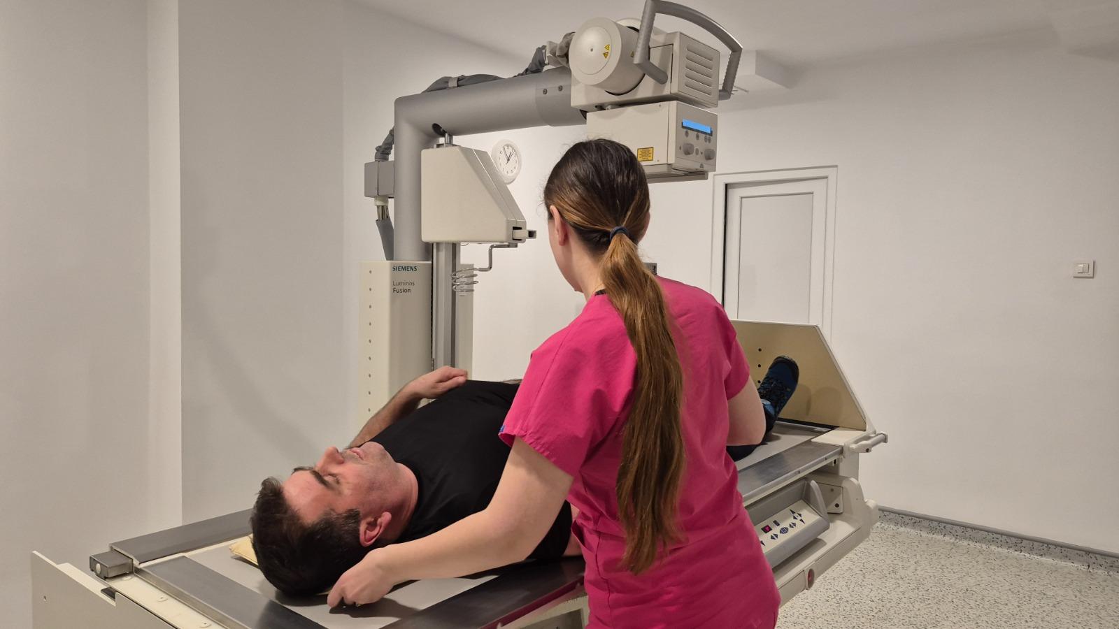
The Procedure
- You'll be positioned by the technologist
- The body part is aligned with the X-ray detector
- You'll be asked to hold still and possibly hold your breath
- The X-ray is taken (takes a fraction of a second)
- Additional views may be taken from different angles
What You'll Experience
- Positioning on the X-ray table or standing against a detector
- Possible use of positioning aids
- Brief moment of stillness during exposure
- No pain or sensation from the X-ray itself
Duration Most X-ray examinations take 5-15 minutes.
After the X-Ray
Results
- Digital images are immediately available to the radiologist
- Formal interpretation typically within hours to next day
- Urgent findings are communicated immediately
- Report is sent to your referring physician
Radiation Safety
We take radiation safety seriously while providing necessary diagnostic imaging.
Understanding X-Ray Radiation
Types of Radiation X-rays use ionizing radiation, which carries some risk. However:
- Modern equipment uses the lowest doses necessary
- Digital technology requires less radiation than film
- Benefits of diagnosis typically far outweigh risks
Dose Perspective Typical X-ray doses are low:
- Chest X-ray: Equivalent to about 10 days of natural background radiation
- Limb X-ray: Very low, equivalent to a few hours of background
- Abdominal X-ray: Slightly higher but still relatively low
Safety Measures
ALARA Principle We follow "As Low As Reasonably Achievable" principles:
- Using lowest dose that produces diagnostic images
- Limiting examinations to those medically necessary
- Shielding sensitive areas when possible
- Using modern, well-maintained equipment
Pregnancy X-rays during pregnancy are avoided when possible due to fetal radiation sensitivity. If X-ray is medically necessary, we use protective measures and the lowest possible dose. Always inform us if you're pregnant or might be pregnant.
Pediatric Considerations Children are more radiation-sensitive. We use special pediatric protocols with reduced doses.
Why Choose Sanconfind for X-Ray Services?
Modern Equipment
Our digital X-ray system provides:
- High-resolution images
- Low radiation doses
- Fast image acquisition
- Reliable, consistent quality
Expert Interpretation
Your X-rays are read by:
- Qualified radiologists
- Using systematic interpretation methods
- With attention to clinical context
- Providing clear, actionable reports
Efficient Service
We offer:
- Minimal wait times
- Quick examination completion
- Rapid result turnaround
- Integration with other services
Comprehensive Capability
Our radiology department handles:
- Routine examinations
- Urgent and emergency X-rays
- Specialized contrast studies
- Follow-up imaging
Frequently Asked Questions
Is X-ray safe?
Yes, when used appropriately. Modern digital X-rays use very low radiation doses. The diagnostic benefit typically far outweighs any minimal risk.
Will I feel the X-ray?
No. You won't feel anything during the X-ray itself. Positioning may be slightly uncomfortable depending on your condition.
How long does an X-ray take?
Most X-rays take just 5-15 minutes. Complex studies or multiple areas take longer.
Do I need to prepare for an X-ray?
Usually no special preparation is needed. Remove metal objects from the area being imaged. Some contrast studies require specific preparation.
Can I have an X-ray if I'm pregnant?
X-rays during pregnancy are generally avoided. If medically necessary, protective measures minimize fetal exposure. Always inform us of possible pregnancy.
When will I get results?
Results are typically available within hours to the next day. Urgent findings are communicated immediately.
Is digital X-ray better than film?
Digital offers numerous advantages: immediate images, lower radiation, image enhancement, easy storage and sharing. It has largely replaced film in modern facilities.
What's the difference between X-ray and CT?
X-ray produces two-dimensional images; CT uses X-rays to create cross-sectional (slice) images. CT shows more detail but uses more radiation. Your doctor recommends the appropriate test.
Schedule Your X-Ray
For quick, accurate radiographic imaging, contact Sanconfind Medical Center.
Contact Us
- Phone: +40 244 990
- Email: office@sanconfind.ro
- Location: Sanconfind Medical Center, Poiana Câmpina, Prahova County, Romania
Our team will schedule your examination promptly and provide any necessary preparation instructions.
Sanconfind Medical Center's digital radiology services combine proven X-ray technology with modern digital capabilities, providing fast, accurate diagnostic imaging with minimal radiation exposure.